Many patients asked me time to time their expectation from acupuncture treatment would be very high just like as the magic needle. There are no magic needles you expect. Everything is going as step by step. Your problem, if you have the chronic conditions, is getting worse during the times. So, the treatments might be needed with the proper time and sessions. That meaning is that you need to be patient.
The musculoskeletal disorders are the quite common disorders at the daily practice. Nowadays, they are frequently seeing those patients in my practice.
Therefore, I’ll discuss and mention those issues as follows for my patients’ references in order to understand their problems. I’ll update the below contents time to time when I have time available.
Repetitive Strain Injury (RSI)
RSI is commonly complaint in the daily practice of my clinic. Commonly, the complaint areas are the wrist, elbow, lower arm where the patients did overuse during the times.
Knee Joint Pain
Knee pain is quite common complaints at the daily practice.
Interestingly, some proportion of the patients are not complaining the pain of knee even if they are not having any cartilage. The recent article mentioned that “But contrary to popular belief, knee osteoarthritis is not caused by (https://pubmed.ncbi.nlm.nih.gov/31192807/) “wear and tear”.” (https://www.sydney.edu.au/news-opinion/news/2024″/08/25/-knee-pain-from-osteoarthritis–what-to-try-instead-of-surgery.html#:~:text=Osteoarthritis%20is%20the%20most%20common,disease%2C%20affecting%202.1%20million%20Australians.&text=Knee%20osteoarthritis%20is%20most%20likely,those%20with%20previous%20knee%20injuries.)
So, most of the patients are having the other soft tissues issues such as tendonitis, ligaments inflammations, joint capsulitis, bursitis, and/or the synovial membrane inflammation.
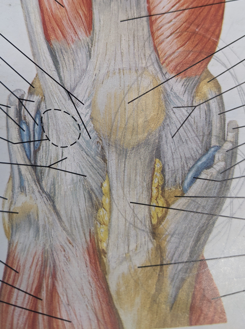
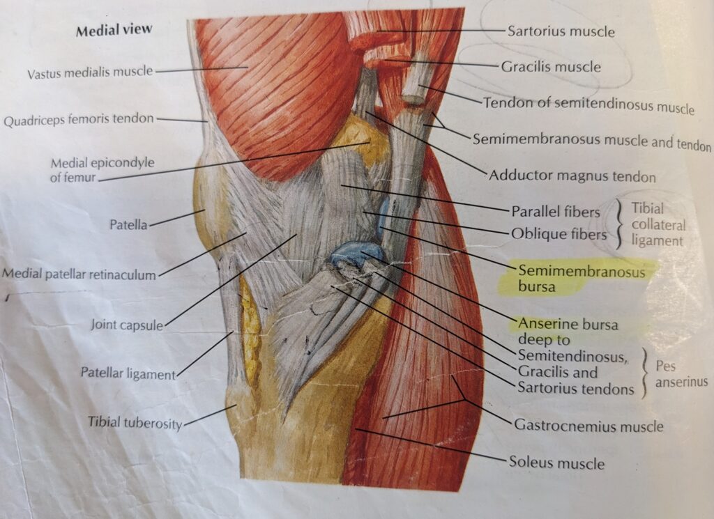
As you can see the above diagram, you can identify the ligaments, joint capsules, bursa, ligaments.
And as you can see the above diagram, you can see the synovial membrane, which is relieving the lubricants of the knee joint. If you have no problem, your knee joint is not swollen, not tight. However, if your knee joint is swollen, heat, redness, and tight, it is showing that you have some inflammation, especially on the synovial membrane which is showing the red lines along the joint capsule.
If you are not treated with those above conditions, your knee joint inflammation is progressing with depositing the bony tissues( Calcium ions) to everywhere inside the knee joint which are called as degeneration, calcification, bony spur formation.
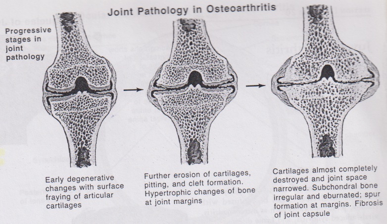
From the above mentioned book, he mentioned that “The most characteristic pathological feature is the growth of osteophytes at the margins of affected joints(nodule or spur formation). The osteophyte, which consists of bone growing from the joint margin, usually follows the contour of the articular surface with the capsule and ligamentous attachments; it may even grow into the joint space and be covered by cartilage that merges into the synovial lining.”
Why are the osteophytes growing?
It is coming from the inflammation of joint capsule, ligaments, and synovial membranes.
For example, you are twisted your knee from down-walking from the hiking. So, you are hurting on the medial collateral ligaments. After you twisted your knee, the medial tibial collateral ligament may be partially teared and it is starting the inflammation reaction such as the pain, heat, swelling, redness. After for a while, the pain may be subsidised and you may feel better. But the pain is not getting away and it looks like up and down. And then suddenly you felt the area you feel pain swollen, lumpy, and/or hard. That is exactly described by nodule or spur formation. It is degeneration processing and you felt weakness, sometimes really sharp pain if the bony spur may be touching the nerve.
Eventually, you decided to see your doctor and then your doctor starts the investigation of your pain by ordering X-ray(if you have any injuries before) and/or CT-scan, MRI scan with Ultra Sound scan. And your doctor may prescribe the pain killer and/or anti-inflammatory medication.
If you came back to your doctor for seeing the results of the X-ray, scans, your doctor may ask you to see the specialist and/or recommend the corticosteroid injection for your pain.
The corticosteroid injection may or may not help you by the radiologist via guiding by CT scan.
Therefore, if your practitioner is able to identify and treat properly, it may help you.
Ligament Inflammation
Many patients are complaining the pain of knee, even if they did surgeries such as Knee Joint Replacements and/or Key hole surgeries. Usually, they were back to their surgeons and discussed with them, and then they got the answer from the doctors without any problems. However, they are still painful.
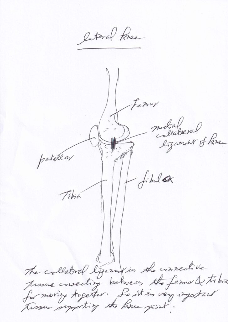
One of the reasons they have the pain of knee is coming from the soft tissues surrounding the knee joint such as the Medial Collateral Ligament of Knee Joint.
As you can see above diagram, the Medial Collateral Ligament of Knee is located at the center of medial knee joint for connection the Femur and Tibia. When you are walking and standing, this ligament is supporting the Knee joint make stabilizing.
With any reasons, if the Medial Collateral Ligament is inflammed with the injuries from the accidents and the overuse, that ligament couldn’t support the knee joint, leading to the pain with difficult walking and standing.
Neck Pain with or without tingling sensation to shoulder/ upper arm/ lower arm/ finger
- Do you have any sore shoulder pain and tingling sensation down to the arm? However, many patients are not mentioning any pain and discomfort at neck. As you can see above diagram, cervical nerves C3, C4, C5, C6, C7, C8, T1 are going down to the shoulder and arm. So, if you are some pains at shoulder and tingling sensation down to the arm, you have to think that you may have some problem at your neck.
- Frequently, I am seeing those patients at the daily practice. When I checked them, they were showing neck muscles were spasmed and inflamed with many reasons.
- Therefore, if you have the problem of your neck, you’ll have the tingling sensation to the arm, weakness, numbness.
- Please don’t ignore. I recommend you have X-ray for your neck and upper back for checking any problems.
- Nowadays, some of patients are showing straight neck from X-ray. Straight neck is called as turtle neck because neck is as like a neck of turtle 🐢. This straight neck is usually coming from wrong posture such as computing, using mobile phone, reading, and work. I’m always saying my patients “chin up”. If you are up your chin, your neck is getting the normal posture and not any more damaging of your neck. Please look after yourself for quality of your life.
Greater Trochanteric Bursitis
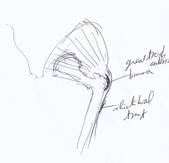
As you can see above diagram, the Greater Trochanteric Bursa is located under Iliotibial Tract above the Greater Trochanter. The Greater Trochanteric Bursa is acting to prevent the fliction between Iliotibial Tract and Bone(Greater Trochanter) as like ball bearing. If there is not Greater Trochanteric Bursa, what is happened? Your tendon which is Iliotibial tract will be torn eventaully after you are using your leg. However, if you are using your body under specification, you don’t have any trouble.
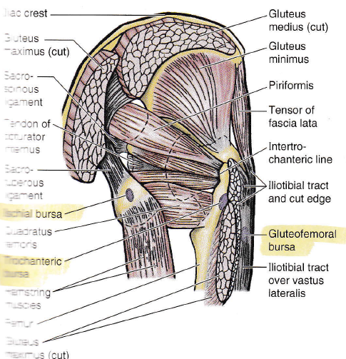
As you can see from the above diagram, trochanteric bursa is not just one. It is located at the lateral greater trochanter and behind greater trochanter. So, above picture is showing the exact location for trochanteric bursa. This bursitis is also quite common disorder in the daily practice.
The patients reported me that they had the corticoid Steroids injection and/or medication such as Endap, which is a brand name for amitriptyline which is a prescription medication, tricyclic antidepressant (TCA) used to treat major depression, chronic nerve pain, migraine prevention, and nocturnal enuresis (bedwetting)(https://en.wikipedia.org/wiki/Amitriptyline#:~:text=Amitriptyline%20is%20indicated%20for%20the%20treatment%20of,than%206%20after%20other%20treatments%20have%20failed.). It works by increasing serotonin and norepinephrine levels in the brain to improve mood and manage pain. However, those treatments are not treating the root problems but just masking the pain. So, when the effect of the medication is vanished during the time, your pain would be coming back. Because the medication may help you that you don’t feel the pain temporally. But how long do you need the medications? Would you like to take the medications for long times?
But we are always making challenge by ourselves to do something such as lots of hiking, lifting heavy objects time to time. For example, if your weight is 60 kgs, and you are lifting and carrying a bag of 20kgs, you are carrying the weight of 80kgs. And furthermore, if you are moving with a bag of 20 kgs, you are actually carrying 2 x 80kg = 160 kgs. It is lots of weights. That meaning is that if you are lifting for a short period of time, it’ll be okay. But if you are doing for a while, your body is strained and eventually your body would be damaged.
How does acupuncture work?
Time to time, even if I explained the mechanism of acupuncture in brochures etc, the patients ask me “How does acupuncture work?”
Please click below link and you’ll get the information you want.
Headace
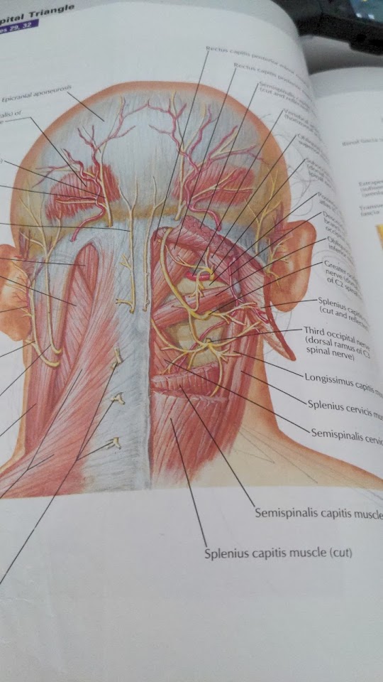
- Cervicogenic headache which is meaning the pain coming from cervical region is quite commonly seeing at the daily practice. Usually, the patients are complaining about the pain of Temporalis muscle. In those patients, they are feeling that the pains are coming from the neck to head. As you can see at the attached picture, the yellow line which is showing the cervical nerves are going from the neck to side of head. Therefore, if your cervical nerves are jammed with any reasons, you may be feeling the pain on head.
- So, there’s many reasons to have nerves jamming with overuse and/or injuries.
Chest Pain
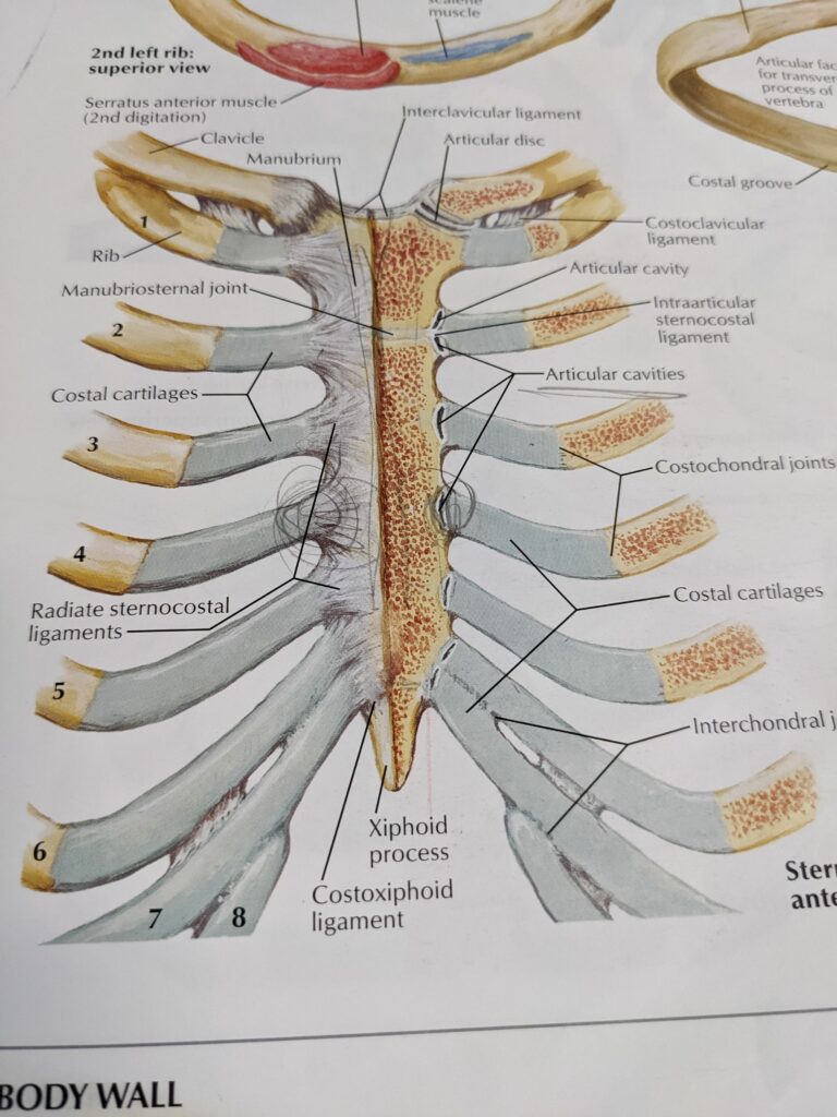
Chest pain is occasionally seeing at daily practice. Many patients are thinking Chest pain may be coming from Heart Attack. Therefore, they are seeing a doctor to investigate the reason why they are feeling the Chest pain. However, after all investigation doctor said that there is no heart problem. Anyhow you need that investigation by a doctor for making sure everything fine. But many patients are still suffering chest pain which may be coming from other reasons. One of the reasons is Sternocostal articular cavities’ problem such as inflammation of radiate sternocostal ligaments, and articular cavities due to injuries of impact accident and/or wrong exercises.
Shoulder Pain
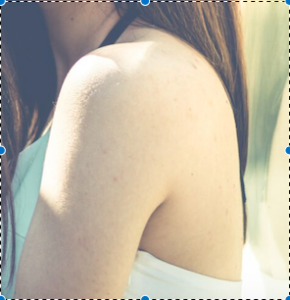
Shoulder pain is quite common. There are two issues such as mobility and pain. Common problem is subacromial bursitis. How do you know whether you have subacromial bursitis? Best option is you visit your doctor by checking your shoulder with ultrasound scan.
– Subacromial Bursitis
Where is subacromial bursa? What is bursa? What function is bursa? How is it happened?
Bursa is water sack structure like as water bump, which is filled with fluids.
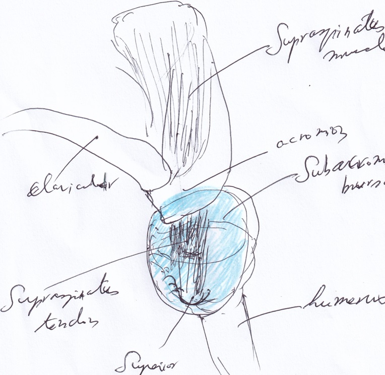
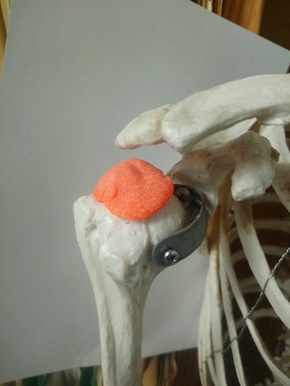
As you can see left side of picture, bursa is located between acromion and head of humerus underneath of supraspinatus tendon.
Bursa is preventing friction between tendon and bone. If tendon is directly touching and moving without bursa, tendon is easily damaged by pricking with bone (head of humerus). Therefore, God designs bursa for preventing direct friction tendon and bone.
Bursitis is meaning inflammation of bursa. Why is bursa inflamed?
The reason is bursa is too much pressured and moving such as abduction by supraspinatus tendon. If Supraspinatus muscle is normal tension, there is no problem to affect bursa. However, supraspinatus muscle is spasmed leading to pulling supraspinatus tendon constantly and moving due to using shoulder.
Signs and symptoms are various. Major signs are pain when you are lying on that side and/or certain movement, swollen, redness, and tight at edge of shoulder. Usually, supraspinatus tendonitis may be occurred with subacromial bursitis. Therefore, you might be concerned both problems for proper treatments.
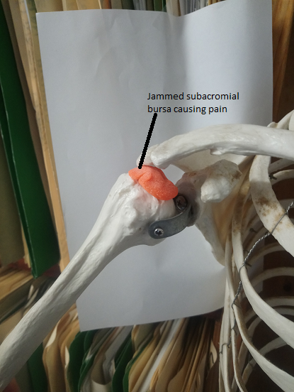
And when you are doing abduction movement like as left picture, swollen bursa is jamming with abduction movement causing pain certain angle between approximately 80 to 120 degrees. So, if you feel pain on shoulder when you are raising your arm above shoulder, that is subacromial bursitis.
Treatments
- Conventional treatments: Doctors might offer you steroid injection at local area. Usually, doctors inject steroid with seeing ultra sound image or X-ray image and prescribe certain medications such as anti-inflammatory and pain killers.
- Acupuncture: We may recommend those patients acupuncture treatments. Because if you are treating by medications and steroid injection, acupuncture doesn’t interfere your conventional treatments from the fact that acupuncture treatments are using only bare needles without any substances.
– Rotator Cuff Tendonitis
Bursitis
Bursitis is inflammation of bursa which is located at several areas of our body. What is bursa doing function?
Bursa has a important function which is preventing friction between bone and tendons and/or ligaments.
Muscle Inflammation
Muscle inflammation is muscle is inflamed due to overuse and any injuries, leading to pain. Muscle inflammation usually came with muscle spasming, tendonitis, and ligament inflammation, furthermore, joint capsulitis, and joint inflammation called as arthritis. Muscle is acting with contraction and relax according to nerve sensation from Central Nerve System.
For example, if you want to grasp a cup, your brain is sending nerve signal to Peripheral Nerve System to specific nerve for controlling muscles which is using for grasping a cup.
However, if you are lifting heavy pots, you might be overusing your muscles. Muscles have a limitation for lifting things such as limitation of rope to pull heavy things. Therefore, muscle fibres are tearing and torn, leading to inflammation of muscle fibres. Inflammations are irritation of sensory nerves which are innervating muscles. Eventually, you’ll feel pain.
Synovitis
Tenosynovitis
Trigger finger

Trigger finger is occasionally seeing in the daily clinic practice. Patients are complaining about discomfort of the finger when they are using the finger. Because when they are extending the finger, they can’t extend with force until certain point and then extend the finger with click noise and pain. However, major patients are eventually they can’t extend the finger. Conventional medicine is injecting steroid and/or surgical operation.
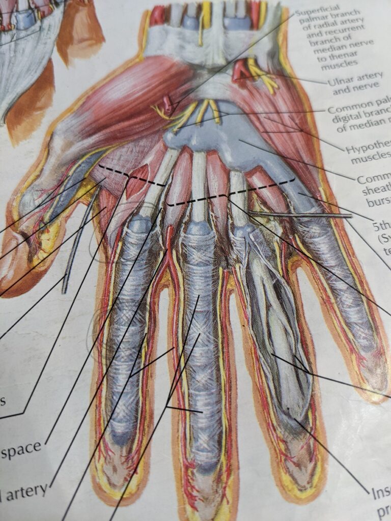
Major problem is inflammation of tendon and tendon sheath and/or anular part of fibrous sheath of Metacarpal joint (MCP) with the reasons, which is one of them is continuous using finger with fixed posture. Tendon sheath which is synovial is functioning for preventing friction between tendon and anular part of fibrous sheath. If tendon sheath is inflamed, synovial tendon sheath is relieving too much synovial fluid, leading to swelling at that area. Eventually swollen tissue can’t move properly at that MCP joint.
Ligament Inflammation
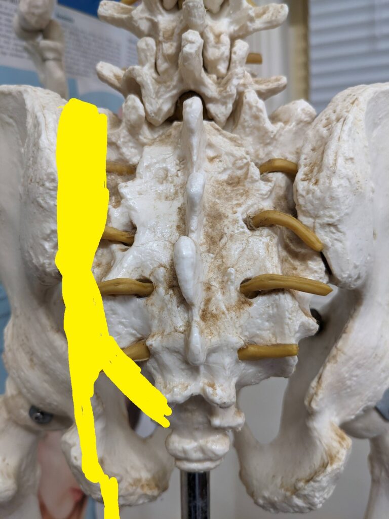
The attached diagram is showing the area of posterior sacroiliac ligaments located at the back side of your hip, which is not showing the muscles but showing the bones(sacrum, iliac). It is quite common presentation in the daily practice. Ligament is very strong tissue which may not be damaged easily. However, any reasons such as overuse, heavy lifting and/or carrying heavy things, sports activities, gym activities, hiking, mountain biking etc could be causing the strained ligaments, leading to the inflammation of ligament. And then those inflammation may make the ligament is getting weakness and fragile with certain activities, even if your daily activities such as walking, standing, sitting, lying down etc. The patients are describing that pain is dull ache and annoying pain continuously. So, you have to concern this problem. Your acupuncture practitioner may help you this problem. Acupuncture treatment may reduce the inflammation, reduce pain, do the healing processing happened. Major issue is that the patients are coming to the clinic at the late stage. So, it may be at the chronic stage. Therefore, it is taking time to resolve all issues with above ligament inflammation. I recommend you that it is better coming earlier than late stage to resolve your issue quickly with less pain and less time.
Sprained Ankle
Sprained ankle is occasionally seeing in daily practice. Most of patients are saying they are injured when they are doing sports activities such as football, netball, and walking with wrong stepping and/or landing holes. Common injury is inversion injury which is lateral foot is going to medial side of foot. When they are injured, there are injured
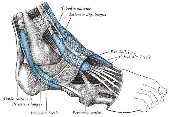
Sprained ankle is quite often seeing at the daily practice. The reasons you had a sprained ankle are many causes. They are miss step on uneven ground, fall with wrong way, etc. However, I recommend you that any sprained ankles need to have attention with X-ray. Because any sprained ankle might be happened with fracture and/or cracks of bones. If you have any fractures and cracks, you need to be medical cares from your doctors. Even though there is no fractures and cracks of bones, many patients are complaining the pain surrounding the ankle. Most of common sprained ankle is the inversion injury. As you can see the attached picture, the inversion injuries are involving several soft tissues which are Extensor Digitorum Longus/Brevis muscle with tendon, Fibularis Longus/Brevis muscles with tendon, Fibularis Tertius tendon, Superior/Inferior Extensor Retinaculum, and Ankle Joint Capsule. Mostly they are over-stretched, tearing. Eventually they are inflamed with signs of swelling, redness, and heat. Acute injury of the sprained ankle is needed to do RICE (Rest, Ice, Compression, and Elevation). After the acute injury if you have still pain at local area, you might have the persistent inflammation that you need to deal with them.
Tendonitis
typical tendonitis is supraspinatus tendonitis, which is annoying dull pain with weakness when doing particular movement by force.
tendonitis has classified as surgical condition to be repaired by surgical operation and nonsurgical conditions not suitable to surgical operation. surgical condition is it might be torn over 1/3 thickness and width of tendon. it could be diagnosed by your doctor with ultrasound scan or MRI. usually doctor repair torn tendon with attaching end of tendon to the bone such as head of humerus. therefore, length of tendon might be shorter than original length of tendon. after surgery, you had rehabilitation physiotherapy for stretching out tendon. anyhow, this stretching can be making tendon longer than torn tendon. however, tendon might be thinner than normal tendon. therefore, tendon is weaker than original tendon. So, you might be understanding weakness than original tendon.
nonsurgical condition is torn area is under 1/3 width and thickness of original tendon. therefore, doctor would like to observe condition what is going on. if the condition is getting worse, doctor wants to offer you surgical operation. if condition is not getting worse, doctor would like to prescribe certain medications such as pain killer and/or anti-inflammatory medications with cortisol injection for under controlling pains.
however, lots of patients are not responding conventional treatments, therefore, they are looking for other modalities such as physio, chiro, osteo, acupuncture, massage. Physio, Chiro, Osteo, Massage are called as manual medicine, which is meaning treatments using hands such as stretching, manipulation, massage.
another nonsurgical condition is micro tear of tendon, leading to tendonitis which is meaning inflammation of tendon. lots of patients are complaining weakness, pain with certain movements as like sometimes sharp usually dull aches, swollen tendon with heat and redness showing typical inflammation. if inflammation is not treated properly, inflammation is going to degeneration, calcification, scar tissue formation leading to tendon is getting thicker, harder, and making lump formation. somebody will think lump formation is tendon is stronger than normal due to thickness than normal size of tendon. however, it is not. hard tissue is not flexible, not elastic. therefore, tendon is easily more damageable than normal tendon.
Achilles Tendonitis
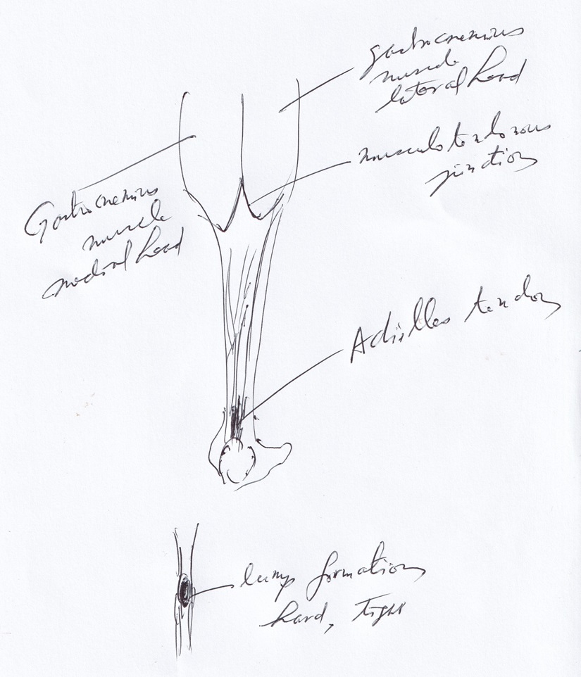
Achilles tendonitis is occasionally seeing at the daily practice. It is coming from running on the beach or farmers who are walking around the muddy ground for a while. It is quite discomfortable disorder, and occasionally seeing the lumps on the Achilles tendon which are showing as above diagram and hard, tender to touch, stiff in the morning, pain and discomfort when walking.
If it is not treated properly, the lump is getting bigger.
What is the lump?
The lump is forming as follows. After the tendon is torn and/or tearing with lots of reason, tendon is inflammed. The inflammation is getting the area of inflammed tendon is swelling, heat, and redness. After time is going on, the area of inflammation is getting harder by the reason of depositing the calcium ions at the local area.
Joint Capsulitis
Upper Back Pain
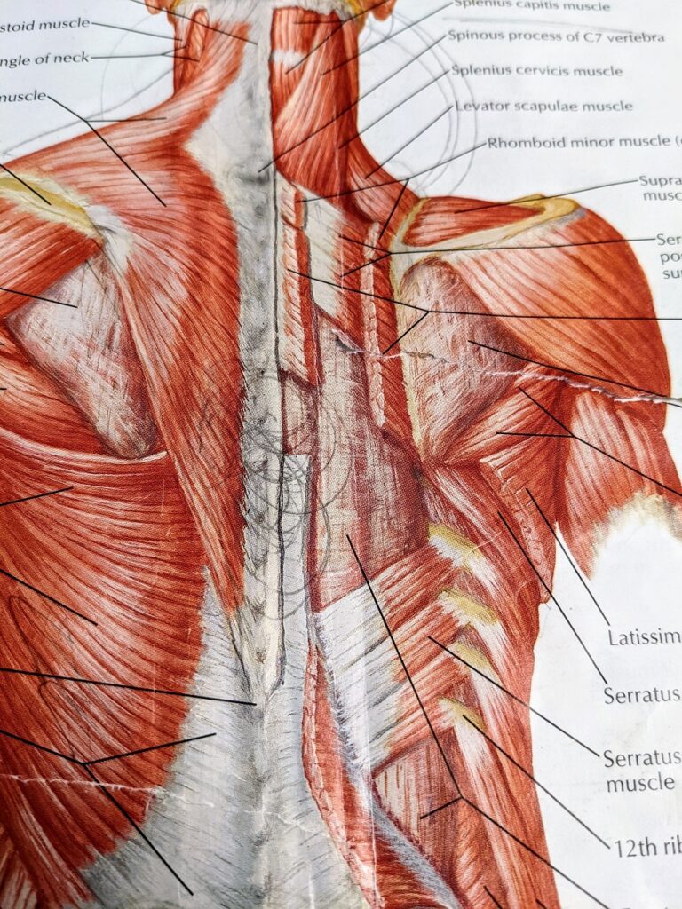
Upper back pain is occasionally seen in daily clinic. Many people are playing sports activities such as rugby, soccer, and fitness. Every sports activity involves the risks of injuries. Especially, lifting and/or scrum while pushing when you are playing rugby are causing strains at the upper back. Typical areas are C7 to T3 spines and Trapezius muscle and Rhomboid minor and major muscles. When those areas are strained, it develops micro injury such as microfibre tearing leading to inflammation of tendons and muscles.
Sometimes you might be developing disc related troubles such as disc bulging out, eventually, leading to tingling sensation and/or shooting down to the arm. They are related with nerve related injuries.
Eventually you would have Tendonitis and muscle inflammation. Those inflammation can irritate the nerves causing pains which are affecting your sleeping. If you feel pain at night making you wake up during the night, it is quite a severe problem you need to solve it out. Please don’t ignore your pain. If you are not treated properly, your problems are getting spread out to other areas. Sports activity is for your recreation and socialise with friends and community. But if you are damaged by those activities, it is not worth it for your daily activities. Looking after yourself.
Nowadays most people are living during 100 years. Take care.
Low Back Pain
Low back pain is quite common disorder.
Hyper lumbar lordosis is as like bow. If string is tight up, it is leading to bow is getting bended. Who is blamed? string.
Bone and muscle
Bones are passive elements, and muscles are active elements for making movement our body. Who are making our body moving? They are muscles. If we don’t have muscles, we couldn’t move at all. Usually, muscles are attached to bones called as origin and insertion.
Pivot mechanism
Cervicogenic Headache
TMJ(Temporomandibular) Disorder
When you feel the pain on jaw, it is quite annoying pain.
Piriformis syndrome
Piriformis
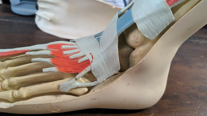
Plantar Fasciitis
Plantar fasciitis is quite frequently seeing one of the common disorders in my daily practice.

As you can see side of photo, our foot is similar structure. Arch is similar to foot bone structure. Bridge is similar to plantar fascia. Arch is supporting bridge weight. Without arch, bridge can’t work. Arch is important structure for bridge. Same principle can be applied to your foot.
If you have a plantar fasciitis, plantar fascia is tearing leading to annoying pain, sometimes bony spur formation causing sharp pain. Major reason is collapsing arch structure of foot.
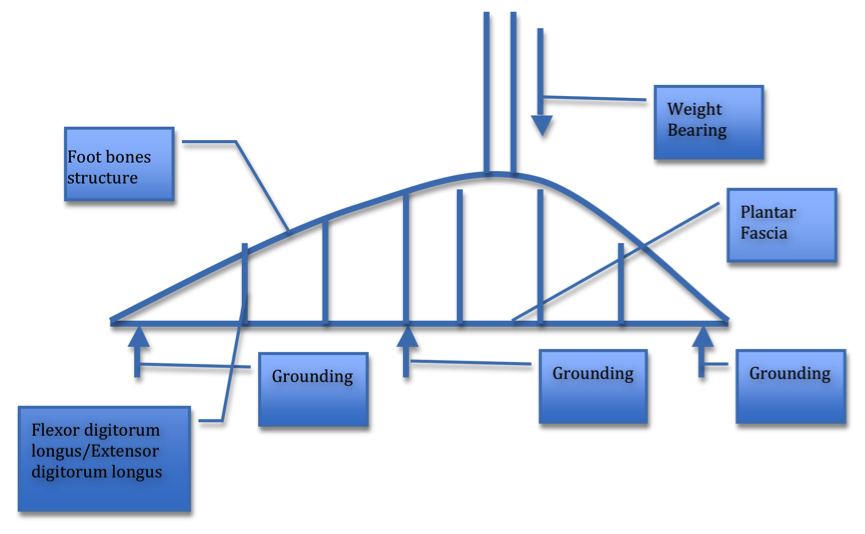
As you can see left side of picture, basic structure supporting weight bearing to foot is foot arch which is foot bone structure that is tarsal bone, hill bone, navicular bone joining with ligament. Furthermore, muscles such as flexor digitorum longus, extensor digitorum longus, etc are supporting arch structure as like bar connecting arch and bridge which is plantar fascia.
Therefore, why is plantar fasciitis developing? As you can see above picture, if bar is broken, arch is collapsing and bridge is also breaking. First of all, bone structure is first thing you have to check with X-ray, CT, and MRI for any fracture. If bone structure is no problem, next one is muscles and tendons to connect between bone and plantar fascia. Definitely, you need to treat plantar fasciitis. If your muscles’ tension are not regulated for examples such as muscle spasms, which is major problem, they might cause plantar fascia tearing, leading to plantar fasciitis (inflammation of plantar fascia).
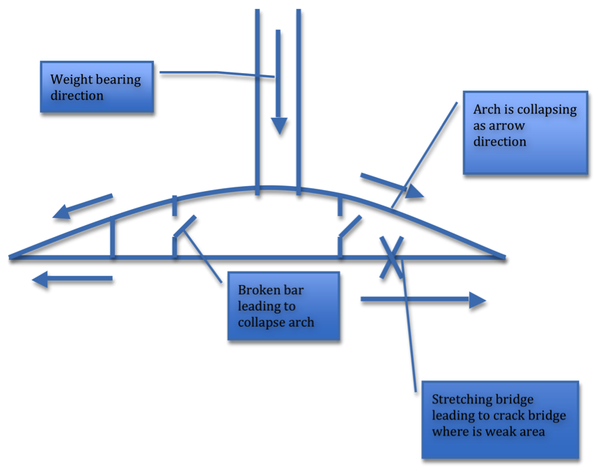
As you can see left side of drawing, broken bar is leading to collapse of arch, further to damage bridge.
How can I fix broken bridge? First of all, you need to fix bar make restoring arch. And finally you can fix bridge.
Same principle may apply to plantar fasciitis.
Therefore, first of all, you need to treat muscles/tendons for restoring arch. And then treat plantar fasciitis.
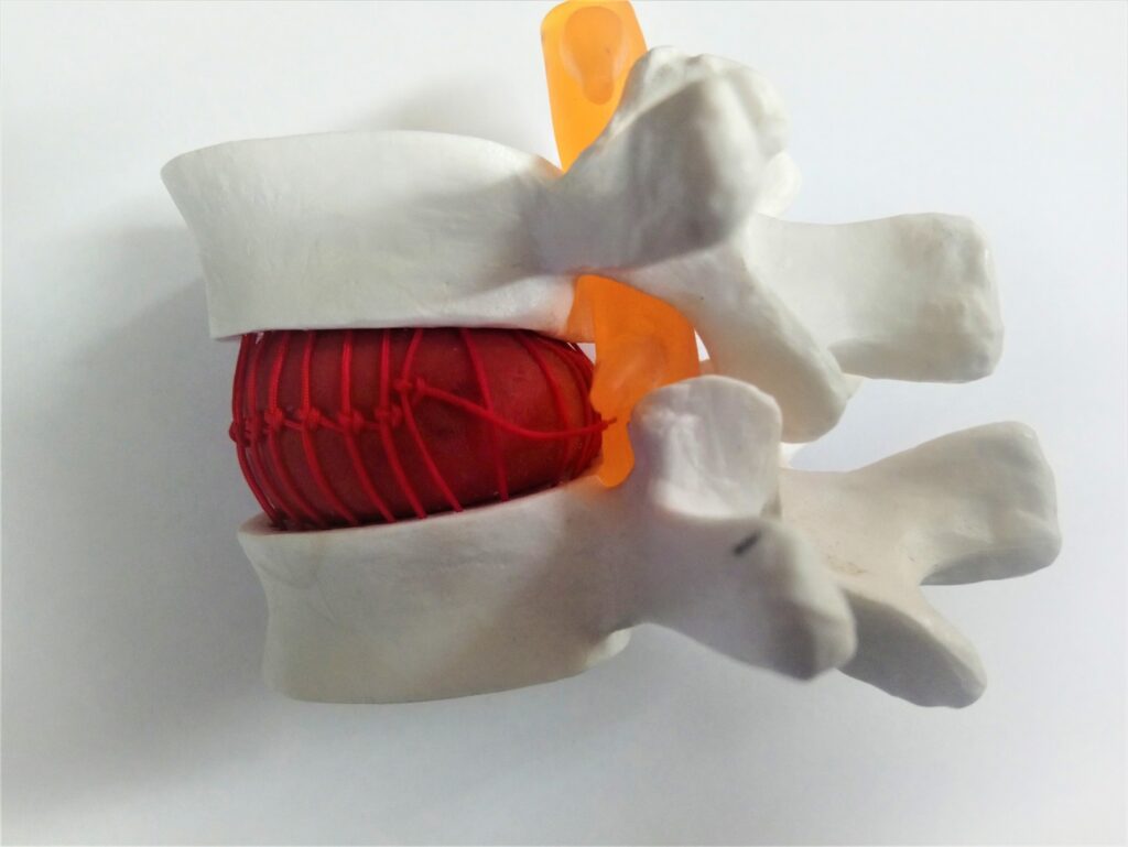
Posterior Sacro-Iliac Joint(SIJ) pain
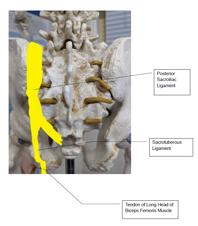
As you can see the above diagram, Ligament is a connective tissue connecting between bone and bone. In this case, the posterior sacroiliac ligament is connecting between Sacrum bone and Iliac bone and working together.